
IV.D. PyMOL Viewing Biological vs. Asymmetric Units
Roderico Acevedo and Kristen Procko
Overview: The activity in this chapter demonstrates different ways to display structures (e.g., biological vs. asymmetric units).
Outcome: The user will explore different tools to load/display information related to biological/asymmetric units.
Time to complete: 10 minutes
Modeling Skill
- Display biological vs. asymmetric units
About the Model
PDB ID: 1hho
Protein: Oxy-hemoglobin
Activity: Gas transport in red blood cells
Description: Tetramer, bound heme prosthetic group with bound oxygen, phosphate ligand
Background
The asymmetric unit of a crystal structure can represent either: (a) the complete biological unit (i.e., the biochemically active form of a biomolecule); (b) a portion of the biological unit; or (c) multiple copies of the biological unit. The “asymmetric unit” is equivalent to the biological unit (correct oligomeric functional state) in approximately 60% of structure records. The RCSB PDB has a detailed page explaining the asymmetric unit, with examples.
CIF Files
By default, PyMOL opens Crystallographic Information File (CIF) with the fetch command, which loads the asymmetric unit of the crystal structure. Sometimes this is the biological unit; however, sometimes this will miss a part of the biological unit—and sometimes it will contain more than one biological unit!
A CIF file can be prompted to load the biological unit by typing each line of the code below. This loads a “two state” model where each state is identical. In the command line, type:
set assembly, 1
fetch 1hho, assembly1, async=0
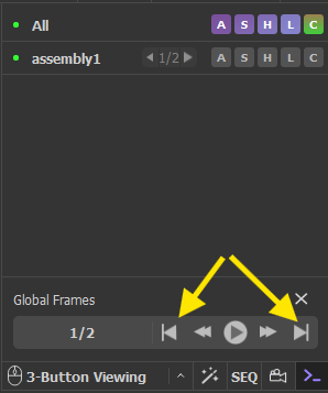
To view each assembly, simply click “next” button in the Global Frames panel (shown below with the yellow arrow).
Experiment with coloring your model by chain (In names/object panel, beside assembly1, click C>By Chain> By Chain).
PDB Files
PDB files are the legacy format for the protein data bank. You can open these with the fetch command as well, and they load the file differently, with each file of the biological unit loading as a separate “state”. The Global Frames panel of the GUI can be used to flip through the states:
fetch 1hho type=pdb1
To view both states simultaneously, the following command can then be executed to create an object for each.
split_state 1hho
Or, the states can be loaded together, as two separate objects. This usually works nicely.
fetch 1hho, type=pdb1, multiplex=1
Going Deeper
About the Models
| PDB ID: 4mqk
Protein: Fetal hemoglobin Activity: Oxygen transport Description: Tetramer (dimer of dimers) |
PDB ID: 6y2e
Protein: SARS CoV-2 main protease Activity: Proteolytic enzyme Description: Dimeric enzyme that activates viral proteins |
Steps
- Load each of these files (one at a time) into PyMOL using the “fetch” command.
- Explore the different assemblies by clicking on them in the log.
What is shown in each assembly? Consider a scenario where you need to examine the biological unit. Consider a scenario when you don’t need the biological unit.
To see the unit cell:
- In the command line, type: show cell
NOTE: You may need to adjust the clipping (use scroll wheel) to see the whole unit cell. If the macromolecule appears outside the cell, the structure may not contain cell data.
Click here to go to Chapter V: Mouse/Trackpad Selection