
XI.C. Mol* Superimposition
Didem Vardar-Ulu and Shane Austin
Overview: This activity demonstrates how to align two objects in the modeling program.
Outcome: The user will be able to superimpose two similar macromolecules to examine commonalities and differences
Time to complete: 10 minutes
Modeling Skills
- Superimposing objects
About the Models
PDB ID: 1xww & 5pnt (different isozymes)
Protein: Low molecular weight protein tyrosine phosphatase
Activity: hydrolyzes Tyr-OPO32- phosphoester bond
Description: single chain, bound SO42- competitive inhibitor, bound glycerol (nonspecific stabilizer)
Steps
Load the Structures: (Figure 1)
-
-
-
-
-
-
-
-
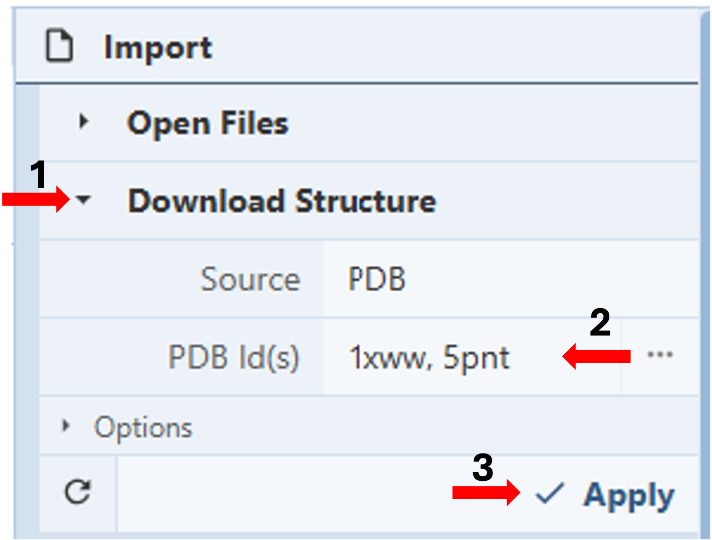
- Go to https://www.rcsb.org/3d-view/ to access an expanded Mol* viewer with an “Import” Panel at the top of the Control Panel.
- Click “Download Structure”. (Arrow 1)
- Type the PDB IDs for the structures you want to superimpose in the cell next to PDB ID(s) under “Download Structures” in the Import Panel, separated by a comma, with or without a space. (e.g., 1xww, 5pnt) (Arrow 2)
- Click “Apply” to display both chains in different colors on the screen (Arrow 3)
-
-
-
-
-
-
-
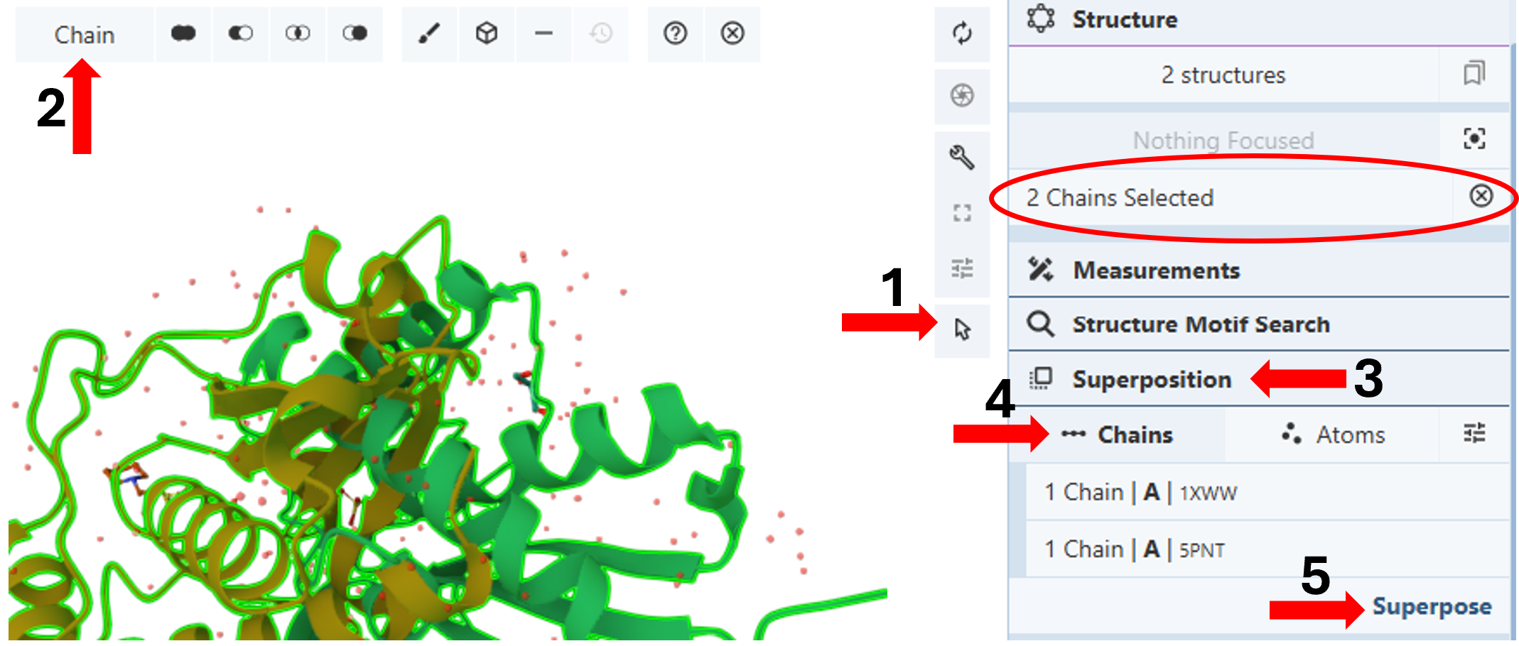
Note: Hovering your cursor on any part of the structure will display information about the PDB ID, model number, instance, chain ID, residue number, and chain name listed at your chosen picking level at the bottom right corner of the 3D canvas.
Superpose by Chains: (Figure 2)
-
-
-
-
-
-
-
-
- Activate the “Select Mode Toggle” by clicking the “cursor” icon. (Arrow 1)
- Choose your picking level as “Chain”. (Arrow 2)
- Click your first chain to select it, either from the 3D Canvas or the Sequence Panel.
-
-
-
-
-
-
-
Note: You will see a green highlight around the selected chain, and the selection will be noted in the “Structure Panel”. (red ellipse)
- Click your second chain to select it.
Note: If you are using the sequence panel, first change the display to the desired protein and chain.
- Click “Superposition”. (Arrow 3)
- Click “Chains”.(Arrow 4)
- Click Superpose. (Arrow 5)
Note: The RMSD between the two chains is reported below the 3D Canvas.
-

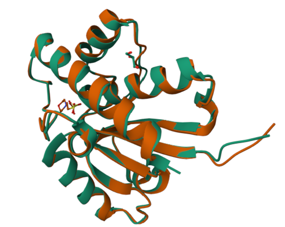
Figure 3: Superposed structure of 1xww and 5pnt Hide the water molecules using the eye icon next to the “water” component.
- Click a white space on the 3D Canvas to deselect chains.
Superpose by Atoms: (Figure 4)
-
-
-
-
-
-
-
-
-
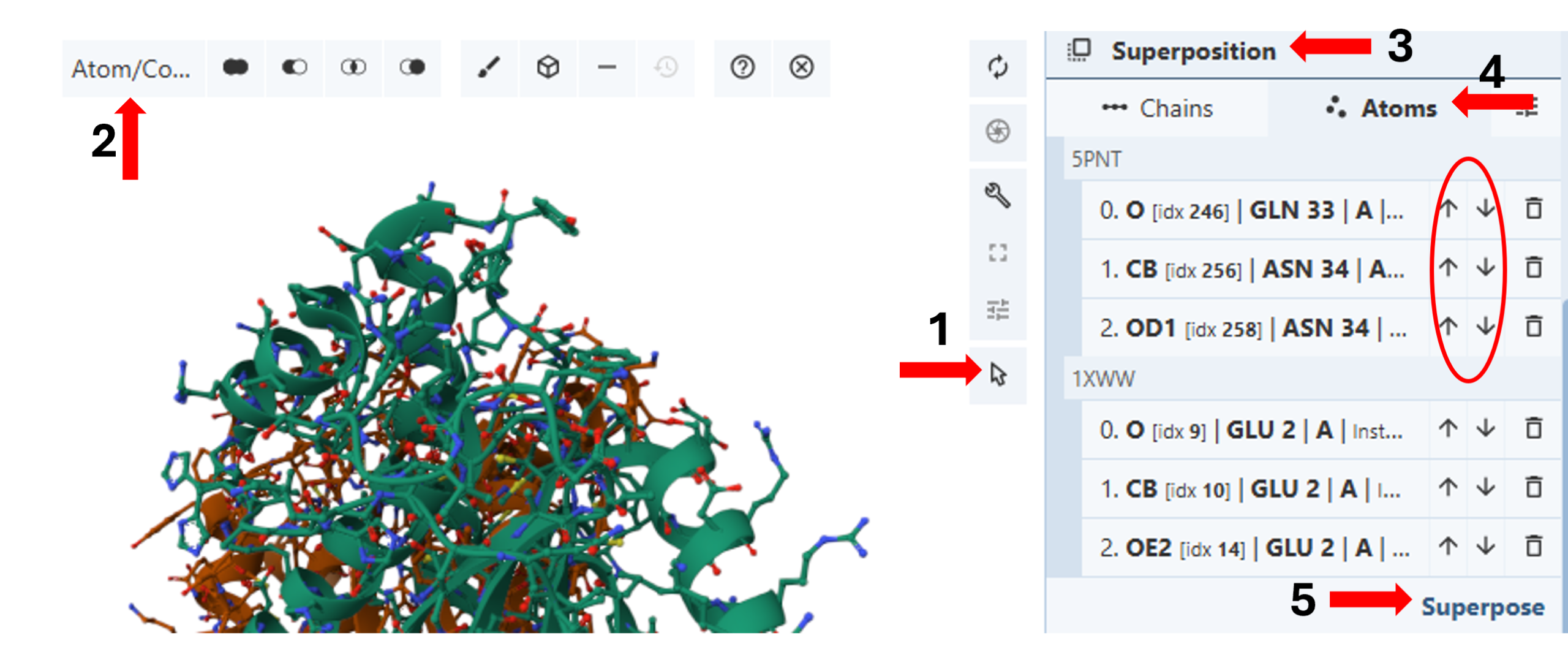
- Create and display ball-and-stick representations for both chains. (see Chapter II.C) “Changing Representation”)
- Activate the “Select Mode Toggle” by clicking the “cursor” icon. (Arrow 1)
- Choose your picking level as “Atom/Coarse Element”. (Arrow 2)
- Click “Superposition”. (Arrow 3)
- Click “Atoms”. (Arrow 4)
- Click one or more atoms you want to include in the superposition from one of the structures.
- Click the corresponding set you want to include in the superposition from the other structure.
- Click Superpose. (Arrow 5)
-
-
-
-
-
-
-
-
Note: The superposition is done on all given pairs of atoms in the order they appear in the superposition panel. To reorder the atoms, use the up and down arrows in the panel. (red ellipse). The RMSD between the two chains is reported below the 3D Canvas.
- Hide the water molecules using the eye icon next to the “water” component.
- Click a white space on the 3D Canvas to deselect chains.