"

XI.B. iCn3D Superimposition

Henry V. Jakubowski and Kristen Procko

Overview: This activity demonstrates how to align two objects in the modeling program.

Outcome: The user will be able to superimpose two similar macromolecules to examine commonalities and differences

Time to complete: 5 minutes

Modeling Skills

  • Superimposing objects

About the Models

PDB ID: 1xww & 5pnt (different isozymes)

Protein: Low molecular weight protein tyrosine phosphatase

Activity:  hydrolyzes Tyr-OPO32-  phosphoester bond

Description:  single chain, bound SO42- competitive inhibitor, bound glycerol (nonspecific stabilizer)

Steps

  1. Open iCn3D
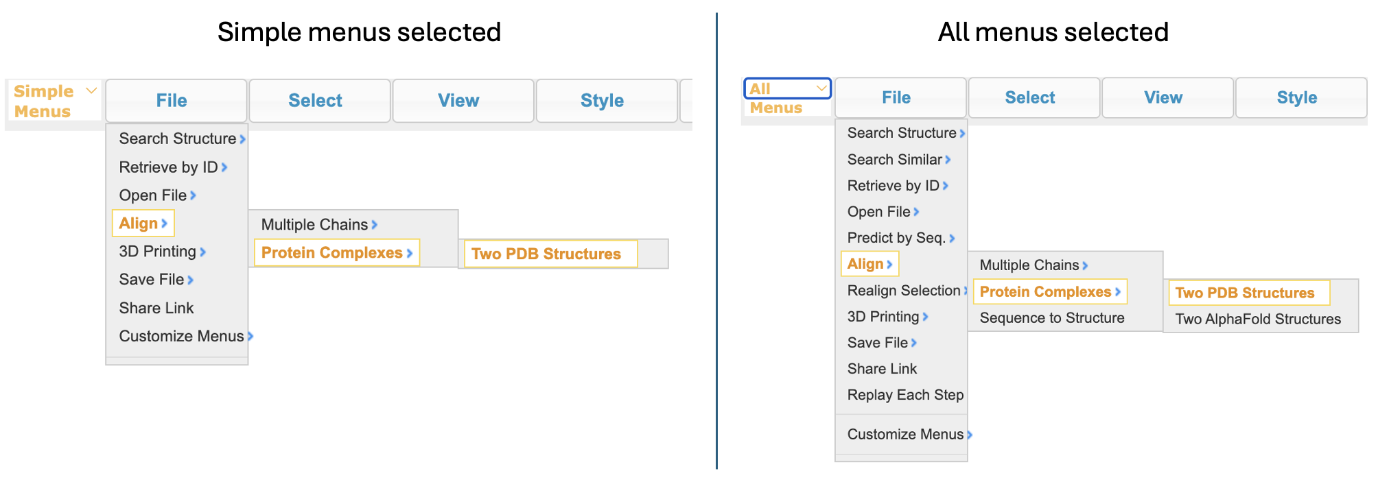
  2. Open and align the two structures. In the dropdown menu: File → Align → Protein Complexes → Two PDB Structures

Note: The menus will have different options depending on whether you have selected “simple menus” or “all menus”.

Screenshots of two menus: Left menu shows the simple file menus with 'File' then ‘align’ then ‘protein complexes’ then ‘Two PDB structures’ highlighted. Right menu show the all menus selected setting with 'file' then ‘align’ then ‘protein complexes’, then ‘Two PDB structures’ highlighted. The right menu has several more features in the file menu compared to the simple menu.
Figure 1: Comparing the align function with simple menus and all menus enabled
  1. In the popup window:
a) For ID1, type: 5PNT
b) For ID2, type: 1NN1
c) Click Invariant Substructures Superimposed
Screenshot of the align two PDB structures menu with yellow arrow indicating the ‘invariant substructure superimposed’ box.
Figure 2: The align structures popup with the invariant substructure superimposed button indicated by a yellow arrow

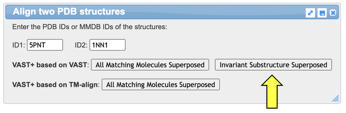
Note: Red regions indicate aligned residues with identical amino acids in the two structures, blue indicates aligned residues but non-identical amino acids, and gray indicates non-aligned residues. See example

Rendering of a protein represented by alpha carbon model in gray (non-aligned), blue (aligned, nonidentical residues), and red (aligned, identical residues) .
Figure 3: Initial output of the dissimilar alignment
  1. Repeat the above steps, this time using 5PNT and 6Y2V, both human phosphatases with 85% sequence ID
  1. Rendering of two aligned proteins in C-alpha trace models with red representing amino acids with conserved identities, blue indicating nonconserved but aligned amino acids and grey representing non-aligned amino acids.
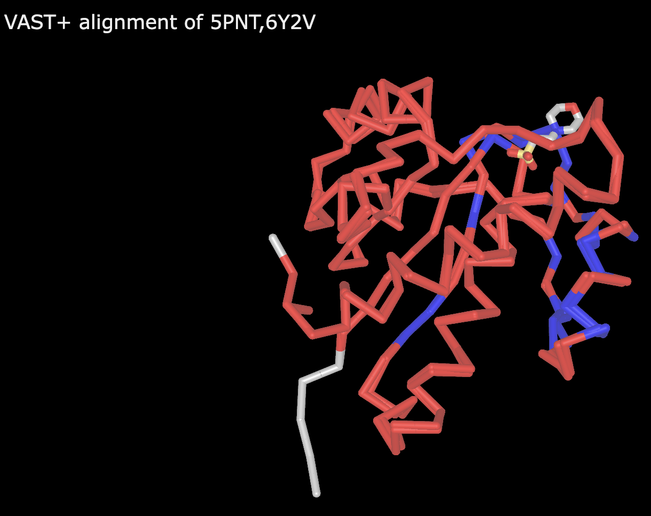
    Figure 4: Alignment of 5PNT and 6Y2V

    With the trackpad or mouse, click anywhere within the structure viewer.

  2. Use the “a” key to toggle between the two aligned structures.


License

Seeing the Invisible: Learning to Teach with Biomolecular Visualization Copyright © by The BioMolViz Working Group. All Rights Reserved.