
I.B. Getting Started with iCn3D
Henry V. Jakubowski and Kristen Procko
Overview: This chapter presents the iCn3D interface, guides the user to load a structure, and introduces trackpad and mouse controls.
Outcome: The user will be able to load a file in iCn3D, perform basic manipulation (zoom, rotate, and center), and save the file as a PNG image and save all work (for iCn3D, this is done by creating a share link).
Time to complete: 5–10 minutes
Modeling Skills
- Load a structure into the modeling program.
- Perform basic structure manipulation (e.g., zoom and rotate) with a trackpad or mouse.
About the Model
PDB ID: 3fgu
Protein: Catalytic complex of Human Glucokinase
Activity: One isoform of the enzyme that phosphorylates glucose in the first step of glycolysis
Description: single chain, with four bound ions and molecules. Potassium ion (K+) is from the crystallization medium and non-essential for biological function. Bound catalytic magnesium (Mg2+). Bound substrate: beta-D-glucose, and bound substrate analog phosphoaminophosphonic acid-adenylate ester (ANP). ANP is a non-hydrolyzable analog of ATP, thus this structure captures a similar view to the catalytic complex (enzyme-substrate complex).
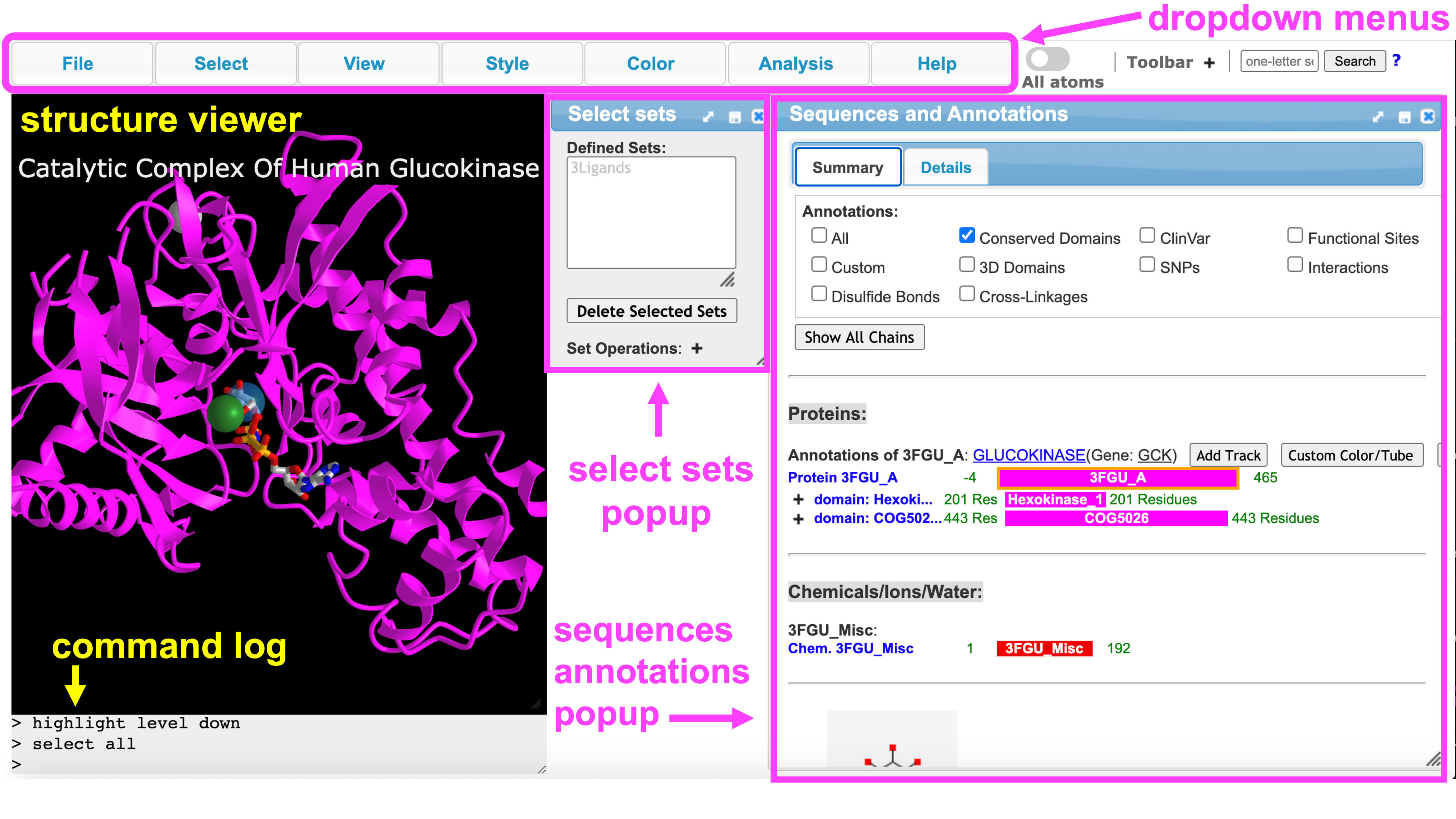
The iCn3D User Interface
Throughout these tutorials, we will refer to the following parts of the interface:
Steps
- Open iCn3D
- In the “List of PDB, MMDB, or AlphaFold UniProt structures” box, type: 3FGU (default is MMDB) Click: Load Biological Unit (for a discussion of Biological vs. Asymmetric Units, see A Guide to PDB Structures and the RCSB PDB)
Note: Default render is ribbon (cartoon) with black background and small molecules shown as sticks
- Now, familiarize yourself with basic structure manipulation using the guide below. Based on your specific trackpad or mouse, these may vary slightly. Play around to see how yours works.
- rotate: click and drag (mouse: left click and drag)
- zoom: pinch and spread (mouse: rotate the scroll wheel)
- translate: two finger click and drag (mouse: right click and drag)
- re-center: left click View from the top menu bar, then select “Center Selection”
Note: ctrl + click on a PC is the same as command + click on Mac
- Position and zoom the structure as desired.
- Save the file in two ways. Using the dropdown menus:
- Save an iCn3D PNG image: a) File → Save Files → iCn3D PNG image → original size
- In the popup window, type a name.
- File can be reloaded in iCn3D: File → Load → iCn3D PNG IMAGE)
- Save an iCn3D share link: File → Share Link → Save → Lifelong Short Url.
- Copy and paste the link into a text file to save it.
Note: The iCn3D PNG link is useful for when you want to save a file to your computer. This can be used as an image in a presentation slide, and saved to open in iCn3D for a class demonstration. The URL is a great tool because you can create a QR code from it that students could scan in class to rapidly pull up the structure on their device. Mainly, what type of file to save depends on instructor preference.
Jump to the next iCn3D tutorial: II.B. iCn3D Alternative Renderings